Proton Calorimetry/Experimental Runs/2021/Apr15
Afternoon test at UCLH with photodiodes and CMOS sesnor.
Experiment Equipment
| Item | Notes |
|---|---|
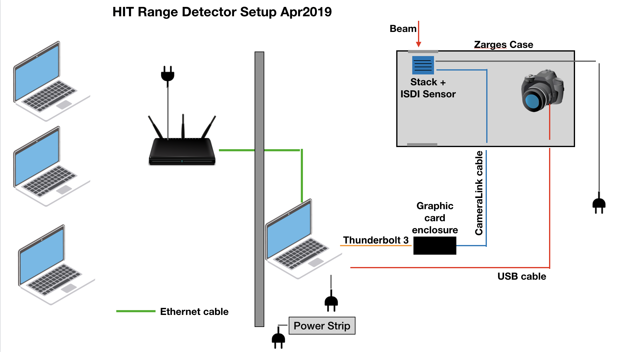
| Network Hub | Set in control room to take output from experimental room ethernet connection. Control laptops connected via 5GHz WiFi. |
| Control Laptop x3 | 2x remote control for sensor and FPGA, 1 for notes/web GUI. |
| Ethernet Cable x 3 | To connect DAQ laptops to network in the experimental room, network hub to network in control room. |
| Portable Enclosure | Modified Big Zarges Waterproof Wheeled Equipment Case.
Features mount for scintillator stack, front and back openings for beam, patch panel with ports for SHV, BNC, SMA, USB, Camera Link cables. |
| Scintillator stack | X x 2 mm, Y x 2.6 mm and Z x 3 mm sheets in ascending order from the front/beam of the end of the scintillator. Sheet numbers from front to end (in beam direction): UNKNOWN. |
| ISDI CMOS sensor | sensor pixel dimension: 1030 x 1536. NO optical grease between scintillator and sensor. Connected to DAQ PC via Camera Link cable. |
| Nikon D7500 | Connected to DAQ Laptop via USB cable. |
| DAQ laptop x2 | Control sensor and photodiode acquisition. |
| Nexys Video FPGA development board. | For interfacing between DDC232 and PC. |
| Rev. 2 Texas Instruments DDC232 custom circuit board (x2) | Each housing 16x Hamamatsu S12915-16R photodiodes, coupled directly to scintillator sheets. |
| Gloves | For handling scintillator |
| Digital Caliper | For measuring PMMA air gaps |
Range Detector Experiment
ISDI Sensor Instructions
General Sensor Configuration
- Base Configuration is 14 x 1 Bit
- Resolution is 1030 x 1536 pixels
- For selecting pixel rows in centre: Use horizontal offset (ignore DVAL to activate field) and horizontal resolution
Increase number of frame buffers
- To increase the number of frame buffers to the allowed maximum, start XCAP as administrator.
- Select Capture/Sequence Capture/Video to Frame Buffers/Driver Assistant
- Tick box in left field, then set number of frame buffers here.
Serial Terminal instructions for ISDI sensor
- Find serial terminal in PIXCI(R) tab of sensor control software.
- In Controls/Options: Make sure "Send string with CR" is selected. Should be default.
- In Controls/Setup: Select Serial Port: Enabled. Baud rate has to be 115200
- Set low full well mode using W000300000
- Set high full well mode using W000300004
- Read full well mode using R0003
Continuous Field buffer setting
- This setting makes sure that the buffer is filled with a new frame as they come in. If this option is not selected the sensor and software will not be synchronised and we *will record a lot of pitch black images.
- Select Capture/Live Options//Live Mode/Live Video: Continuous All buffer capture
Experiment Plan
12–13th April
- Find convenient beam intensity that does not saturate sensor: lowest clinical beam energy, smallest beam focus
- Calibration of detector (highest clinical energy). Shoot from front and back of stack (we have a new entrance window at the back of the stack). Also calibrate at different focus sizes. Also test camera: focus on front of beam stack and centre of beam stack and compare.
- Test focus (beam size) dependence: shoot pencil beam with ~10 cm range at different focus sizes
- Measure WET of known blocks of plastic to calibrate range telescope using a pencil beam with ~10cm range. Lennart will check if he can find some PMMA slabs whose WET has been accurately measured. Ideally have some slabs between ~1mm and ~5cm WET thickness. We know that there are some PMMA slabs between 1mm and 5cm thickness, measured by Giulia Arico (OMA fellow). These measurements will be used to benchmark Laurent's reconstruction code. Use tightest beam focus.
- Repeat 0.-3. with all available ions (protons, helium, carbon and oxgen)
- Measure SOBP of protons. 10cm deep, 3cm plateau
- Measure high proton energies (up to 250MeV) using PMMA absorber in front of range telescope.
- Measure a couple of oxygen pencil beams (range shift) and SOBP
- Disassemble detector.
13–14th April
- Shoot-through calibration for Helium and Carbon: 430.1MeV/u Carbon 30cm range, He 220.5MeV/u 30cm range.
- High full-well mode C Intensity: 8*10^7; He Intensity: 8x10^8.
- Low full well mode C Intensity: 3*10^7; He Intensity 3*10^8.
- Rotate detector; repeat.
- Carbon and Helium SOBP and single energy pencil beam:
- 2 Gy SOBP Carbon, Helium mix keep same intensity here and scale down later in data processing.
- Carbon and Helium Intensities both 8*10^7 (low Helium intensity!). Both high and low full well modes. Only measure low full well as sensor starts to come out of saturation.
- Carbon SOBP max range ~9cm (plateau 3cm longitudinal, 3x3cm2 lateral), He SOBP max range ~27 cm.
- Repeat with 220MeV/u pencil beam (C 10cm, He 30 cm).
- Add PMMA blocks, repeat previous measurement. 24 measurements; 5 changes in room. (5+5+5+2+2cm PMMA blocks, 19cm PMMA in total). Both high and low full well mode.
- Variable air gap measurements:
- 5cm PMMA, air gap, remaining PMMA absorber; total thickness of 20.8cm. Downstream sheets 5+5+2cm PMMA sheets plus half of unused pairs.
- Vary air gap width: 0mm, 2mm, 5mm, 10mm.
- Vary air gap depth: 1mm, 2mm, 5mm, 10mm.
- Upstream PMMA absorber 5cm; downstream PMMA absorber varies from 12.8cm to 13.7cm.
- 32 measurements, 16 absorber changes.
- For 5mm x 5mm gap, adjust focal spot size.
- Energy 220 MeV/u for both; intensity 8*10^7 for both; use focus setting 3 (8.5mm for Carbon; 8.1mm for Helium).
- Low full well mode for sensor.
- Half covered 2mm air gap:
- 5cm upstream PMMA, 2mm air gap on left-hand side, 13.6cm downstream PMMA absorber. 18.8cm total PMMA thickness.
- Sheet pulled out, 8mm pulled out from centre, half-centred, 8mm pushed in from centred, all the way in.
- Energy 210 MeV/u for both; intensity 8*10^7 for both; use focus setting 3 (8.5mm for Carbon; 8.1mm for Helium).
- Scan layer with 10x10 spots over 3x3cm area.
- Low full well mode for sensor.
- Take movie with DSLR.
- Moving platform measurements:
- Assemble PMMA stack on moving platform: upstream PMMA absorber 5cm; 5mm air gap; downstream PMMA absorber 13.3cm. Total thickness still 18.8cm.
- Gap width 5mm; gap depth 5mm.
- Irradiate with single energy Helium plan: 10x1 spot array (3cm wide) and move platform at the same time. Spot dwell time ~200ms. Total plan irradiation time: 5s. Total platform movement: 4cm.
- Take images with sensor: use 100 pixel wide sensor strip and long acquisition.
- Energy 210 MeV/u for both; intensity 8*10^7 for both; use focus setting 3 (8.5mm for Carbon; 8.1mm for Helium).
- Lung phantom measurements only(!) if enough time. Take lung phantom, place it on height adjustable table, place tumor in beam (180 MeV/u C and He), lift it by 0.5 cm
14–15th April
- Shoot-through calibration for Helium and Carbon: 430.1MeV/u Carbon 30cm range, focus 4.9mm; He 220.5MeV/u 30cm range, focus 3.4mm. 5cm upstream PMMA absorber.
- High full-well mode C Intensity: 8*10^7; He Intensity: 8x10^8.
- Low full well mode C Intensity: 3*10^7; He Intensity 3*10^8.
- Lung phantom measurements with tumour.
- Align lung phantom with lasers. Lung tumour (about 1cm^3) sits positioned at isocentre.
- Monoenergetic pencil beam: Helium energy 180 MeV/u, Intensity 8*10^7, spot focus 3, Carbon 180MeV/u, intensity 8*10^7, spot focus 3.
- Use optimal full well mode for sensor: low full well mode.
- Step phantom vertically:
- 9 different heights.
- 5 mm vertical spacing.
- Start 20 mm below tumour, step phantom downwards until beam 20 mm above tumour.
- 8 changes in room plus initial setup; 18 total measurements (Carbon+Helium for each position).
- High energy Helium Bragg peak scan with absorber.
- Switch to higher Helium energies (remote controlled by accelerator crew):
- Carbon: energy 324.26 MeV/u, intensity 8*10^7, spot size 8mm.
- Helium: energy 324.26 MeV/u, intensity 7*10^8, spot size 7mm.
- Detector measurements in both high and low full well mode.
- Build up 50 cm of PMMA in front of detector. Remove PMMA stack in 10cm steps: final measurement with 10cm, 5cm and 0cm absorber in place to give shoot-through calibration.
- 7 PMMA steps, Carbon+Helium, high+low full well mode: 28 total measurements.
- After first measurement with 50cm WET absorber, measure activation of PMMA and detector with full stack in place.
- Switch to higher Helium energies (remote controlled by accelerator crew):
- ADAM PETer phantom measurements.
- Use sensor high and low full well modes.
- Continue with higher Helium energies:
- Carbon: energy 324.26 MeV/u, intensity 8*10^7, spot size 8mm.
- Helium: energy 324.26 MeV/u, intensity 7*10^8, spot size 7mm.
- Setup: Place phantom in upright position. Fill with oil (measurements need to be fast, phantom heavily leaking oil). Place 2cm PMMA degrader in front of phantom; 15cm PMMA absorber between phantom and detector. Energy degraded down to ~300 MeV/u at phantom. Upstream face of Zarges case 28cm from isocentre.
- Align ADAM PETer in isocenter:
- Vertical shift by 5 mm.
- Start at -1cm and step upwards.
- 5 positions, 10 measurements, 4 changes in room.
- Align ADAM PETer in isocenter, rotate:
- 4 measurements (0, 2, 4, 10 degree).
- 4 measurements, 3 changes in room plus setup.
- ADAM phantom measurements.
- Use optimal sensor full well mode: probably high full well.
- Continue with higher Helium energies:
- Carbon: energy 324.26 MeV/u, intensity 8*10^7, spot size 8mm.
- Helium: energy 324.26 MeV/u, intensity 7*10^8, spot size 7mm.
- Setup: Place ADAM in isocentre. 15cm PMMA absorber between phantom and detector.
- 5 measurements: Uninflated rectum, 30 Ml Air, 45 Ml Air, 60 Ml Air, 75 Ml air. Rectum ballon inserted to 15cm.
- 5 measurements, 4 changes in room.
- Align isocenter with predefined spot. Repeat previous measurements. 5 measurements, 4 changes in room.
- Repeat measurement with spot in between isocentre and predefined rectum spot (horizontal alignment isocenter, vertical 1 cm below isocenter), and spot above isocenter (horizontal alignment isocenter, vertical alignment 1cm above).
- Repeat measurement with ADAM rotation:
- Align ADAM in isocenter.
- Rotate by 2 degree steps.
- Repeat measurement with ADAM horizontal shift:
- Align ADAM in isocenter.
- Horizontal shift by 1 cm.
- ADAM lower energy phantom measurements with AP beam (30 degree gantry rotation).
- Use optimal sensor full well mode: probably high full well.
- Switch to lower Helium energies:
- Carbon: energy 263.4 MeV/u, intensity 8*10^7, spot size 8mm.
- Helium: energy 263.4 MeV/u, intensity 7*10^8, spot size 7mm.
- Setup: place ADAM in isocentre in upright position.
- Repeat rectal balloon and rotation measurements.
- Water equivalent thickness measurements of 10 x 1cm sheets:
- Use optimal sensor full well mode: probably high full well.
- Shoot-through calibration with 220 MeV (30cm) proton range; 5cm upstream absorber.
- Switch to monoenergetic proton beam with 10cm range.
- Insert sheets one at a time and measure range shift.
- 11 measurements, 10 setup changes in room.
Experiment Notes
Range calorimeter measurements with ISDI sensor + scintillator stack.
12–13th April
images from sensor: User/Public/Document/EPIX/XCAP/data/20190512
images from dslr: User/Public/Document/Nikon/
- full sensor area
- shooting on BACK WINDOW
- Simon checking the intensity on pixels from dslr with nikon software
- camera settings
- exposure 1/2 sec
- aperture f/3
- ISO 200
| Run number | Full Well Mode | Beam Energy (MeV/u) | Range (estimated) | N particles (Hz) | Spot size (mm) | ion type | DSLR file name | Degrader WET (mm) | Comments | Thumbnail |
|---|---|---|---|---|---|---|---|---|---|---|
| N | well mode | Energy MeV/u | Range | particles | Spot FWHM | test | dslr file name DSC_ | degrader WET | comments | |
| 00 | low | None | - | None | None | - | - | 0 | background | |
| 01 | high | None | - | None | None | - | - | 0 | background | |
| not saved from Laurent | high | None | - | None | None | - | DSC_145 | 0 | intensity check | |
| none | - | - | - | - | - | - | - | 0 | Calibration : shoot through. no degrader | |
| 02 | high | 221.06 | - | 2*10^9 | 8.1 | p | DSC_147 | 0 | shoot through no degrader | |
| 03 | high | 221.06 | - | 3.2*10^9 | 11.1 | p | DSC_149 | 0 | shoot through no degrader. from now on exposure time on dslr 0.2 sec | |
| 04 | high | 221.06 | - | 3.2*10^9 | 20.0 | p | DSC_150 | 0 | shoot through no degrader. | |
| 05 | low | 221.06 | - | 1.2*10^9 | 20.0 | p | DSC_151 | 0 | shoot through no degrader. exposure time back to 0.5 sec | |
| 06 | low | 221.06 | - | 1.2*10^9 | 11.1 | p | DSC_152 | 0 | shoot through no degrader. | |
| 07 | low | 221.06 | - | 1.2*10^9 | 8.1 | p | DSC_153 | 0 | shoot through no degrader. | |
| 08 | high | 220.51 | - | 8*10^8 | 4.9 | He | DSC_155 | 0 | shoot through no degrader. exposure 0.1 sec | |
| 09 | high | 220.51 | - | 8*10^8 | 8.1 | He | DSC_156 | 0 | shoot through no degrader. | |
| 10 | high | 220.51 | - | 8*10^8 | 10.1 | He | DSC_157 | 0 | shoot through no degrader. | |
| 11 | high | 430.1 | - | 8*10^7 | 3.4 | C | DSC_159 | 0 | shoot through no degrader. ripple filter 3mm | |
| 12 | high | 430.1 | - | 8*10^7 | 7.8 | C | DSC_160 | 0 | shoot through no degrader. ripple filter 3mm | |
| 13 | high | 430.1 | - | 8*10^7 | 20 | C | DSC_161 | 0 | shoot through no degrader.ripple filter 3mm. exposure 0.5 sec | |
| 14 | high | 430.1 | - | 8*10^7 | 3.4 | C | DSC_162 | 59 | shoot through. PMMA 5 cm | |
| 15 | high | 430.1 | - | 8*10^7 | 7.8 | C | DSC_163 | 59 | shoot through. PMMA 5cm | |
| 16 | high | 430.1 | - | 8*10^7 | 20 | C | DSC_164 | 59 | shoot through. PMMA 5cm | |
| 17 | high | 220.51 | - | 8*10^8 | 4.9 | He | DSC_165 | 59 | shoot through. PMMA 5 cm. exposure 0.1 | |
| 18 | high | 220.51 | - | 8*10^8 | 8.1 | He | DSC_166 | 59 | shoot through. PMMA 5cm | |
| 19 | high | 220.51 | - | 8*10^8 | 10.1 | He | DSC_167 | 59 | shoot through. PMMA 5cm | |
| 20 | high | 221.06 | - | 3.2*10^9 | 8.1 | p | DSC_168 | 59 | shoot through. PMMA 5 cm. exposure 0.2 | |
| 21 | high | 221.06 | - | 3.2*10^9 | 11.1 | p | DSC_169 | 59 | shoot through. PMMA 5cm | |
| 22 | high | 221.06 | - | 3.2*10^9 | 20 | p | DSC_170 | 59 | shoot through. PMMA 5cm | |
| 23 | high | 430.1 | - | 8*10^7 | 3.4 | C | DSC_172 | 0 | shoot through. no absorber. exposure 0.1 | |
| 24 | high | 430.1 | - | 8*10^7 | 7.8 | C | DSC_173 | 0 | shoot through. no absorber | |
| 25 | high | 430.1 | - | 8*10^7 | 20 | C | DSC_175 | 0 | shoot through. no absorber. exposure 0.2 sec |
