Proton Calorimetry/Experimental Runs/2019/Apr12-15
UPDATE THIS PAGE 2 night shifts with range calorimeter and single PMT module.
Equipment List
| Item | Notes |
|---|---|
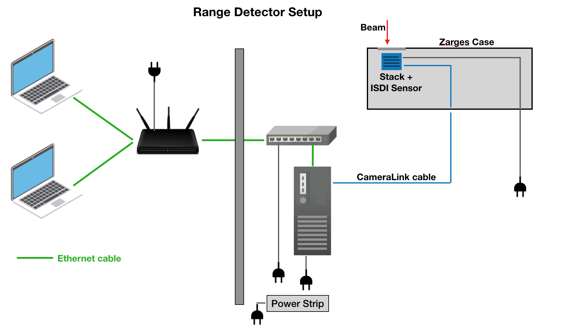
| Network Hub | Set in control room to take output from experimental room ethernet switcher. Control laptops connected via ethernet or 5GHz WiFi. |
| Control Laptop x2 | |
| DAQ desktop PC | |
| Ethernet Switcher | Set in experiment room and connected DAQ desktop PC. Output sent to control room Network Hub. |
| Ethernet Cable x 5 | To connect DAQ PC to switcher, switcher to Network Hub (long cable), 2 laptops to Network Hub, if scope: scope to switcher. |
Range Detector Experiment
| Item | Notes |
|---|---|
| Portable Enclosure | Modified Big Zarges Waterproof Wheeled Equipment Case.
Features mount for scintillator and PMT, opening for beam, and ports for SHV, BNC, SMA, Camera Link cables. |
| Scintillator stack | 14 x 2 mm, 15 x 2.6 mm and 20 x 3 mm sheets in ascending order from the front/beam of the end of the scintillator. Sheet 21 not used. Sheet numbers from front to end (in beam direction): 30,29,28,27,26,22,19,18,17,15,14,13,10,7,25,24,23,20,16,12,11,9,8,6,5,4,3,2,1,31,32,33,34,35,36,37,38,39,40,41,42,43,44,45,46,47,48,49,50. |
| ISDI CMOS sensor | sensor pixel dimension: 1030 x 1536. NO optical grease between scintillator and sensor. Connected to DAQ PC via Camera Link cable. |
| DAQ desktop PC | Controls sensor aquisition. |
| Gloves | For handling scintillator |
Experiment List
12–13th April
0. Find convenient beam intensity that does not saturate sensor: lowest clinical beam energy, smallest beam focus
1. Calibration of detector (highest clinical energy). Shoot from front and back of stack (we have a new entrance window at the back of the stack). Also calibrate at different focus sizes. Also test camera: focus on front of beam stack and centre of beam stack and compare.
2. Test focus (beam size) dependence: shoot pencil beam with ~10 cm range at different focus sizes
3. Measure WET of known blocks of plastic to calibrate range telescope using a pencil beam with ~10cm range. Lennart will check if he can find some PMMA slabs whose WET has been accurately measured. Ideally have some slabs between ~1mm and ~5cm WET thickness. We know that there are some PMMA slabs between 1mm and 5cm thickness, measured by Giulia Arico (OMA fellow). These measurements will be used to benchmark Laurent's reconstruction code. Use tightest beam focus.
4. Repeat 0.-3. with all available ions (protons, helium, carbon and oxgen)
5. Measure SOBP of protons. 10cm deep, 3cm plateau
6. Measure high proton energies (up to 250MeV) using PMMA absorber in front of range telescope.
7. Measure a couple of oxygen pencil beams (range shift) and SOBP
8. Disassemble detector.
13–14th April
0. Calibration for Carbon and calibration for helium at high full well mode. Also test reproducibility: Measure same pencil beam before and after re-assembling detector.
1. Measure Helium and Carbon SOBP through full range of both helium and Carbon. Similar as last time use real SOBP here. The SOBP measurement is useful to show what we are looking at. Measurements taken in step of 5cm PMMA slabs, as last time. Measure in high full well mode of detector. Beam intensities fixed to optimal output of detector. Check source switching time.
2. Repeat 1. with pencil beams but using the realistic beam intensities (2x10^8 for C12 and 2x10^7 for helium). Keep high full well mode for this (measurement useful as reference for intensity scaling of 1 later on).
2.5 Calibration for low full well mode (all following should be in that mode for highest accuracy on carbon fragments)
3. Repeat 2 but in low full well mode for best possible accuracy for helium beam and Carbon fragments. If signal gives too low resolution, find a good helium intensity for onward tests.
4. Use single pencil beam at highest energy (220MeV/u for largest fragment build up) and build small air gaps in the center of the "target": what is the smallest WET change we can see despite the Carbon fragments? What is the smallest lateral gap size we can see for each gap depth? Measure different beam focus sizes. At what depth should the air gap be placed?
->Test 4 different WET differences: 1mm, 2mm, 0.5cm, 1cm. Laterally, Lennart has to check what is the smallest PMMA slab they have two of. Use that to change the gap lateral size.
5. Potentially: Repeat 4 for lower energy. (could be left out)
6. Repeat 4 with different beam intensities (1 higher, 1 lower intensity). Question to be answered: how much helium do we need for this detector? Not needed if we use unrealistic beam intensities following measurement 2. Could also be left out and done in data processing. (not important for us)
7. Build an online moving air gap that was visible well in 4. Laurent has to set up a quick analysis to judge whether the change is visible. For this, build the PMMA slabs on top of the moving platform available at HIT. Lennart will prepare the remote controlling of the moving platform. Irradiate SOBP Plan and move platform across. Take a video of the central pixel signal. We could repeat this even and take a screen-video as we see the online changes when we take the full sensor in integral mode. Can use DSLR camera for this as well. Might be problematic because we can only see the horizontal movement from the side, i.e. we cannot make use of the focus of the camera. We should still be able to see a range mixing of parts of the beam going through the air gap and parts of it going through the full PMMA phantom.
8. Repeat 7 step-wise (6 steps of 0.5cm depending on the time available) and take full view of CMOS for both helium and Carbon. This sequence of images can be combined to a film (gif) in offline analysis.
9. Repeat 7 and 8 for the simple lung phantom. Interesting for publication and Joao has especially requested these measurements. Clarify what Joao is expecting to see. Movie of moving bragg curve with phantom an moving platform?
Measurement 4 and onward could be performed with realistic and/or unrealistic high helium intensity. The latter would give more resolution, but less realism. Lennart would say, that we see in 3 if our signal quality is good enough with the realistic beam and then decide what to do. We can always use high intensities and scale the signal down in data processing. Laurent believes that a helium intensity of 2*10^7 p/s will be observable in our detector but might be a faint signal. We need to decide on the fly if the resolution is ok or not. If not, use higher helium intensity.
14–15th April
0. Shoot-through calibration with 324 MeV/u Helium and Carbon beam (we can also use standard calibration energies, i.e. the highest clinical energies for helium and carbon), helium will be at an intensity of 7*10^8 (that is what was pre-set by the HIT engineers) it can be lowered if needed, but Lennart is still finding out how controlled that is (i.e if we get feedback on the intensity from the accelerator of if we are "blind").
1. Take full depth-dose curve of Carbon and helium. This would be an interesting test to see the signal from Helium at these high energies. Lennart is not sure if he will find enough PMMA with accurately measured WET but is working on it (we need 50 cm PMMA roughly).
2. Shoot through ADAM and measure both Helium and Carbon tail after Phantom. Aim at prostate close to rectum. Lennart chose the 324MeV/u according to a treatment plan. This carbon beam will stop in the prostate close to rectum.
3. Blow up the rectal balloon (2 steps: 50ml air, 75ml air), irradiate both helium and carbon. Do we see changes?
4. Reposition beam/rectal balloon if no change is visible. repeat 2 and 3 as needed.
5. Use no rectal balloon, aim at Prostate and fill Bladder in 2 steps: Do we see changes (no drastic changes expected)? Do only if plenty of time!
6. Aim at center of prostate and rotate phantom in 2 degree steps. Do we see changes? Some issue here is the hull of the phantom, which has a high gradient (curvature and dense material), so changes in range could be related mostly to that and the test might not be very realistic. Can also move phantom laterally as this measurement was recommended by Markus Stock from MedAustron. Could also be left out if under time pressure. Please be more specific about details of measurement: Alignment of phantom
7. Place the phantom vertically and aim at prostate. Measure both helium and carbon. Blow up rectum again (2 steps again) and show difference in range.
8. If still time/energy resources: more realistic lung phantom measurements. Lennart has yet to prepare a plan for these, as he only got the phantom recently. Alternatively, we could investigate the simple lung phantom from GSI, if we weren't able to do that in the second night shift.
9. There is a new pelvis phantom with fixed prostate available at HIT. It can be used for the measurement of horizontal displacements.
Experiment Log
PMMA blocks are 50mm thick. Water Equivalent Thickness is 57.81mm.
Beam Tests
12–13th April
Beam focus ranges from step 1 (smallest) to step 4 (of 6).
Helium ion beam
full sensor area
| Run number | Full Well Mode | Beam Energy (MeV/u) | Range (estimated) | N particles | Spot size (mm) | Test type | Comments | Thumbnail |
|---|---|---|---|---|---|---|---|---|
| N | well mode | Energy MeV/u | Range | particles | Spot FWHM | test | comments | |
| 00 | None | None | - | None | None | - | background | |
| 01 | low | 50.57 | 22.9 | 10^8 | 18.6 | He | low energy, low intensity |
Carbon ion beam
full sensor
| Run number | Full Well Mode | Beam Energy (MeV/u) | Range (estimated) | N particles | Spot size (mm) | Test type | Comments | Thumbnail |
|---|---|---|---|---|---|---|---|---|
| N | well mode | Energy MeV/u | Range | particles | Spot FWHM | test | comments | |
| 44 | high | None | - | None | None | - | background |
Proton beam
full sensor
| Run number | Full Well Mode | Beam Energy (MeV/u) | Range (estimated) | N particles | Spot size (mm) | Test type | Comments | Thumbnail |
|---|---|---|---|---|---|---|---|---|
| N | well mode | Energy MeV/u | Range | particles | Spot FWHM | test | comments | |
| 79 | high | None | - | None | None | - | background |
14–15th April
Beam focus ranges from step 1 (smallest) to step 4 (of 6).
Helium ion beam
full sensor area
| Run number | Full Well Mode | Beam Energy (MeV/u) | Range (estimated) | N ions /s | Spot size (mm) | Test type | Comments | Thumbnail |
|---|---|---|---|---|---|---|---|---|
| N | well mode | Energy MeV/u | Range | ions/s | Spot FWHM | test | comments | |
| 00 | high | None | - | None | None | - | background | |
| 01 | high | 50.57 | 22.9 | 8x10^8 | 18.6 | He | low energy, max clinical intensity |
Mixed Helium and Carbon ions beam
full sensor
| Run number | Full Well Mode | Beam Energy (MeV/u) | Range (estimated) | N particles | Spot size (mm) | Test type | Comments | Thumbnail |
|---|---|---|---|---|---|---|---|---|
| N | well mode | Energy MeV/u | Range | particles | Spot FWHM | test | comments | |
| 09 | high | 182.32 - 222.31 | 74.4 - 104.5 | - | 5.5 - 4.7 | C | Real Clinical SOBP, 6 pixel rows (512 offset), 100mm distal edge using clinical weighting with approx. equal time to deliver each layer with single on-axis spot. |
