Background/Cell Biology And Division: Difference between revisions
SimonJolly (talk | contribs) No edit summary |
SimonJolly (talk | contribs) No edit summary |
||
| Line 55: | Line 55: | ||
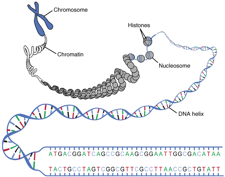
<br />Figure 4: DNA wrapping to form a chromosome [https://en.wikipedia.org/wiki/String_(computer_science)]. | <br />Figure 4: DNA wrapping to form a chromosome [https://en.wikipedia.org/wiki/String_(computer_science)]. | ||
</div> | </div> | ||
DNA is stored in the cells in structures referred to as chromatin. | |||
DNA is wrapped around 8 histone proteins to form a nucleosome. | |||
Tightly bound collections of nucleosomes are referred to as chromatin. | |||
During cell division, these structures condense to form chromosomes. | |||
Healthy humans have 23 chromosomal pairs (or 46 chromosomes). | |||
== Common Terminology used in Genetics == | |||
The following list of definitions commonly used in genetics | |||
; Genes : Different sections of DNA control very specific functions. A gene is a section of DNA that encodes a particular protein (which in turn, regulates a particular function). | |||
; Allele : Human cells contain 2 copies of every gene: one from the mother and one from the father. An allele is a single copy of a gene. For example, a gene may control the colour of the human iris. One allele may cause the iris to be coloured blue whereas another allele may cause the iris to be coloured green. | |||
; Recessive : a recessive allele is one that is only expressed if both alleles are the same. For example, if the allele for green eyes was recessive, the eye colour would only be green if both alleles (copies of the gene) encoded green eyes. | |||
; Dominant : a dominant allele is one that is always expressed. | |||
; Co-dominant : sometimes, both alleles are dominant but encode different proteins. For example, blood type can be classified as either A or B. Some individuals will have AB blood, indicating that both the A allele and the B-allele are being expressed. | |||
== Cell Division == | |||
<div style="text-align: center;"> | |||
[https://en.wikipedia.org/wiki/File:Cell_Cycle_2-2.svg https://upload.wikimedia.org/wikipedia/commons/thumb/e/e0/Cell_Cycle_2-2.svg/587px-Cell_Cycle_2-2.svg.png] | |||
<br />Figure 5: Schematic of the cell cycle [https://en.wikipedia.org/wiki/Cell_cycle]. | |||
</div> | |||
The rate of cell division is highly variable: different cell-groups will divide at different rates. | |||
There are two main types of cell division: mitosis and meiosis. | |||
Somatic cells (i.e: those not involved in sexual reproduction) divide by mitosis. | |||
Germ cells (those involved in sexual reproduction) primarily divide by meiosis. | |||
Figure 6 effectively summarises the cell cycle for both mitosis and meiosis (both of which are indicated by the letter M). Prior to cell division, cells are in a state that is termed interphase (I). Interphase itself can be subdivided into 3 components: G1, S and G2. Cells that are not dividing or in interphase are said to be senescent and exist in the “G0 phase”. Cells may enter the G0 phase for long time periods, or indefinitely, but can re-enter the cell cycle if the correct biological signal is received. | Figure 6 effectively summarises the cell cycle for both mitosis and meiosis (both of which are indicated by the letter M). Prior to cell division, cells are in a state that is termed interphase (I). Interphase itself can be subdivided into 3 components: G1, S and G2. Cells that are not dividing or in interphase are said to be senescent and exist in the “G0 phase”. Cells may enter the G0 phase for long time periods, or indefinitely, but can re-enter the cell cycle if the correct biological signal is received. | ||
During G1 (gap or growth phase) cells increase in size, produce proteins for DNA replication and increase the number of organelles (such as mitochondria). During S-phase (synthesis phase), DNA replication occurs; i.e: the total DNA contents of the cell doubles (however the cell-size does not change). This is illustrated in Figure 7 in which individual the chromatids of the chromosomes (red and blue) are replicated. After replication each chromosome has 2 chromatids (but the total number of chromosomes is unchanged). The two chromatids are connected centrally by a centromere. | During G1 (gap or growth phase) cells increase in size, produce proteins for DNA replication and increase the number of organelles (such as mitochondria). During S-phase (synthesis phase), DNA replication occurs; i.e: the total DNA contents of the cell doubles (however the cell-size does not change). This is illustrated in Figure 7 in which individual the chromatids of the chromosomes (red and blue) are replicated. After replication each chromosome has 2 chromatids (but the total number of chromosomes is unchanged). The two chromatids are connected centrally by a centromere. | ||
| Line 94: | Line 109: | ||
# Image taken from: [https://en.wikipedia.org/wiki/DNA_base_flipping https://en.wikipedia.org/wiki/DNA_base_flipping] (accessed: 04/07/2017). | # Image taken from: [https://en.wikipedia.org/wiki/DNA_base_flipping https://en.wikipedia.org/wiki/DNA_base_flipping] (accessed: 04/07/2017). | ||
# Image taken from: [https://en.wikipedia.org/wiki/String_(computer_science) https://en.wikipedia.org/wiki/String_(computer_science)] (accessed: 04/07/2017). | # Image taken from: [https://en.wikipedia.org/wiki/String_(computer_science) https://en.wikipedia.org/wiki/String_(computer_science)] (accessed: 04/07/2017). | ||
# Image taken from: [https://en.wikipedia.org/wiki/Cell_cycle https://en.wikipedia.org/wiki/Cell_cycle] (accessed: 04/07/2017). | |||
Revision as of 15:22, 4 July 2017
The Cell
In order to understand how cancer develops, it is first important to understand the biology of the cell. The cell is the most fundamental structure to all living organisms: it is an autonomous unit that makes up all organisms and is capable of self-replication. Cells exist in two types: prokaryotic and eukaryotic. All human (and plant) cells are classed as eukaryotic, whereas bacterial cells are categorised as prokaryotic. Although there are many similarities between these two cell types, the main difference is the presence of a membranous envelope around the nucleus of eukaryotic cells.

Figure 1: Eukaryotic cell [1].
Eukaryotic cells are composed of a variety of different structures (termed organelles) including a nucleus, mitochondria, ribosomes and a cell membrane (Figure 1). This list is not exhaustive, but is sufficient to understand the basic biology of cancer. The mitochondrion (sg.) or mitochondria (pl.) of the cell function to convert energy in the form of glucose into one that is usable by the cell. Ribosomes help synthesise proteins. The cell membrane controls the entry and exit of substances (including drugs) into and out of the cell. The nucleus houses the genetic makeup of the cell (the DNA — deoxyribonucleic acid). The nucleolus resides within the nucleus and functions to produce ribosomal RNA (rRNA) which is important for the synthesis of proteins. It is important to realise that the composition of eukaryotic cells varies widely such that a blood cell and heart cell, for example, will have radically different microscopic appearances.
The Nucleus
In eukaryotic cells, the nucleus contains almost all of the genetic material of the cell in the form of DNA (deoxyribonucleic acid). DNA is a double-stranded molecule that is composed entirely of carbon, hydrogen, nitrogen, oxygen and phosphorus. Its’ estimated length is 3 metres (ca.). DNA contains the instructions for cell growth, cell division and cell functionality. Although different cell-classes (e.g: blood cells, heart cells) will express different sections of DNA, the DNA contents of all biological cells, in a health individual, is identical.
Structure of DNA
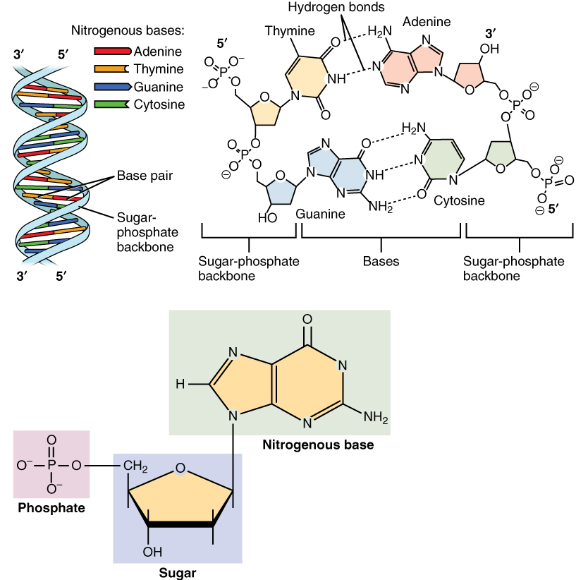
Figure 2: Nucleotides [2].
Figure 3: DNA structure [3].
DNA is composed of two poly-nucleotide stands, wrapped around each other to form a double helix (Figure 3). The term poly-nucleotide refers to the fact that DNA is composed of two string of thousands of nucleotides. Nucleotides are molecules composed of a nitrogenous base, a deoxyribose sugar molecule and a phosphate group (Figure 2). There are 4 nitrogenous bases: cytosine (C), guanine (G), adenine (A) and thymine (T) (Figure 3). Bases on one DNA strand will pair with a base on the second strand. Adenine will always pair with thymine and cytosine will always pair with guanine.
Figure 4: DNA wrapping to form a chromosome [4].
DNA is stored in the cells in structures referred to as chromatin. DNA is wrapped around 8 histone proteins to form a nucleosome. Tightly bound collections of nucleosomes are referred to as chromatin. During cell division, these structures condense to form chromosomes. Healthy humans have 23 chromosomal pairs (or 46 chromosomes).
Common Terminology used in Genetics
The following list of definitions commonly used in genetics
- Genes
- Different sections of DNA control very specific functions. A gene is a section of DNA that encodes a particular protein (which in turn, regulates a particular function).
- Allele
- Human cells contain 2 copies of every gene: one from the mother and one from the father. An allele is a single copy of a gene. For example, a gene may control the colour of the human iris. One allele may cause the iris to be coloured blue whereas another allele may cause the iris to be coloured green.
- Recessive
- a recessive allele is one that is only expressed if both alleles are the same. For example, if the allele for green eyes was recessive, the eye colour would only be green if both alleles (copies of the gene) encoded green eyes.
- Dominant
- a dominant allele is one that is always expressed.
- Co-dominant
- sometimes, both alleles are dominant but encode different proteins. For example, blood type can be classified as either A or B. Some individuals will have AB blood, indicating that both the A allele and the B-allele are being expressed.
Cell Division
Figure 5: Schematic of the cell cycle [5].
The rate of cell division is highly variable: different cell-groups will divide at different rates. There are two main types of cell division: mitosis and meiosis. Somatic cells (i.e: those not involved in sexual reproduction) divide by mitosis. Germ cells (those involved in sexual reproduction) primarily divide by meiosis.
Figure 6 effectively summarises the cell cycle for both mitosis and meiosis (both of which are indicated by the letter M). Prior to cell division, cells are in a state that is termed interphase (I). Interphase itself can be subdivided into 3 components: G1, S and G2. Cells that are not dividing or in interphase are said to be senescent and exist in the “G0 phase”. Cells may enter the G0 phase for long time periods, or indefinitely, but can re-enter the cell cycle if the correct biological signal is received. During G1 (gap or growth phase) cells increase in size, produce proteins for DNA replication and increase the number of organelles (such as mitochondria). During S-phase (synthesis phase), DNA replication occurs; i.e: the total DNA contents of the cell doubles (however the cell-size does not change). This is illustrated in Figure 7 in which individual the chromatids of the chromosomes (red and blue) are replicated. After replication each chromosome has 2 chromatids (but the total number of chromosomes is unchanged). The two chromatids are connected centrally by a centromere. DNA replication ensures that each daughter DNA molecule is identical to the parent DNA molecule. This is achieved by splitting the parent DNA molecule into its constituent strands. The enzyme that catalyses this biological process is termed DNA helicase. The enzyme DNA polymerase is then involved in the assembly of a new DNA strand. This processed is summarised in the following schematic. For simplicity the two strands are shown parallel to each other (rather than being coiled around each other).
In the G2 phase (second gap / growth period) cells continue to grow in size. Proteins required for cell division are primarily produced in the G2 phase, including microtubules (which make up a variety of structures including spindle fibres).
Cell division
As mentioned above, cells can divide by mitosis or meiosis. Both mitosis and meiosis are sub-divided into prophase, metaphase, anaphase and telophase. Mitosis consists of a single cell division whereas meiosis consists of two cell divisions.
Mitosis:
In prophase the nuclear envelope breaks down, the chromatin supercoils to form chromosomes and spindle fibres form. In metaphase, the spindle fibres attach to the centre of the chromosomes and cause them to line up in the centre of the cell (show in Figure 9). In anaphase, the arms of the chromosomes are pulled towards opposite ends of the cell (now shown in figure). In telophase, the spindle fibres break down. The cell then divides into two separate, genetically identical entities in a process called cytokinesis.
Meiosis:
In meiosis, the prophase – anaphase cycle happens twice. In prophase I the nuclear envelopes break down, chromatin condenses to form chromosomes and the spindle fibres form. Chromosomes with the same genes (but potentially different alleles) – also called homologous chromosomes - will pair up and swap genetic material. Thus a chromosome (1a) encoding for blue eyes, and brown hair (for example) could swap genetic material with a chromosome (1b) encoding for green eyes and black hair. After genetic switching, chromosome 1a may encode for blue eyes and black hair and chromosome 1b may encode green eyes and brown hair. This genetic switching encourages genetic diversity in future generations. In metaphase I, homologous chromosomes line up in the centre of the cell and the spindle fibres attach to the individual chromosomes. The chromosomes are then separated into two separate cells during anaphase I, telophase I and cytokinesis I. The spindle fibres degenerate. Prophase II – cytokinesis II resembles mitosis, in which individual chromatids (arms of the chromosomes) are separated into individual cells. The result of meiosis is that the total genetic content of the daughter nuclei at the end of meiosis II is half that of the genetic content of the original cell, prior to interphase.
References
- Image taken from: https://en.wikipedia.org/wiki/Cell_(biology) (accessed: 14/06/2016).
- Image taken from: https://en.wikipedia.org/wiki/Nucleotide (accessed: 04/07/2017).
- Image taken from: https://en.wikipedia.org/wiki/DNA_base_flipping (accessed: 04/07/2017).
- Image taken from: https://en.wikipedia.org/wiki/String_(computer_science) (accessed: 04/07/2017).
- Image taken from: https://en.wikipedia.org/wiki/Cell_cycle (accessed: 04/07/2017).


